Surgical Therapy
Lasers can be prohibitively expensive, and a laser surgeon should carefully calculate the return on the investment. Some of the Er:YAG systems currently available include the Contour dual-mode (ablation and coagulation) laser (Sciton; Palo Alto, Calif), the carbonate (CO3) variable-pulsed laser (Cynosure; Chelmsford, Mass), the CB Erbium/2.94 uniform beam laser (HOYA ConBio; Fremont, Calif), the Venus-i uniform beam laser (Laserscope; San Jose, Calif), and the NaturaLase Erbium laser (Focus Medical; Bethel, Conn).
Some Er:YAG laser systems are no longer available because their manufacturers have concentrated on carbon dioxide or nonablative lasers. The cost of the lasers ranges from $50,000-80,000. Many companies, including third-party vendors, lease the systems for $1,000-1,500/mo with a long-term lease or approximately $600 for 4 hours without a lease. Preowned systems are also available for purchase.
Preoperative Details
Antiviral prophylaxis against cutaneous herpesvirus infection is essential before any full-face or perioral resurfacing; the epidermal trauma and thermal effects often reactivate labial herpes simplex virus. Because of the high prevalence of latent virus, even in persons who have never been symptomatic, prophylaxis is administered to all patients, beginning 24 hours before the procedure and continuing for 7-10 days. Famciclovir (250 mg) or valacyclovir (500 mg) twice daily are effective regimens. Those with a history of herpesvirus infection may receive 500 mg twice daily. [32, 33]
Patients are instructed to avoid taking aspirin or other nonsteroidal anti-inflammatory medicines for at least 10 days in an effort to decrease bleeding during the procedure.
Pretreatment of the skin before laser resurfacing is controversial. No consensus has been reached regarding the usefulness of any particular agent or the usefulness of the practice at all. Most laser surgeons prescribe topical retinoic acids, hydroquinone bleaching agents, and alpha-hydroxy acids for several weeks preoperatively to reduce postoperative inflammation and hyperpigmentation. A typical 6-week preparation regimen before laser resurfacing is as follows:
-
Retinoic acid cream (0.05-0.1%): Apply 0.5-1 g in the evening.
-
Hydroquinone cream (2-4%): Apply 0.5-1 g twice daily.
-
Alpha-hydroxy acid cream (2-4%): Apply 0.5-1 g in the morning.
Studies have not shown these regimens to have a significant effect; hydroquinone is cytotoxic to melanocytes and inhibits tyrosinase, thereby decreasing melanosome formation, but it probably does not reach the deeper melanocytes responsible for hyperpigmentation. Postoperative treatment of hyperpigmentation is more successful. [1, 34] Some advocate the use of antihistamines such as loratadine 30 minutes before using a high-energy Er:YAG laser in an effort to prevent immediate urticarial edema. [35]
Intraoperative Details
If general anesthesia from a concomitant procedure is not used, anesthesia is normally performed by injecting lidocaine mixed with epinephrine. Intravenous sedation is generally required. Topical anesthesia can be used in those procedures in which the Er:YAG laser is only applied superficially to a small area, although the hemostatic effect of epinephrine is lost.
The goal of Er:YAG laser resurfacing is the controlled, layer-by-layer ablation of skin while avoiding the creation of thermal injury so deep that scarring or other complications result. [1] Three variables are adjusted on the laser to create the desired tissue effect: spot size, fluence, and pulse repetition rate. Many laser surgeons use a 3- to 5-mm spot size, a pulse energy of 1-2 J, and a pulse repetition rate of 1-10 Hz. A fluence of 5 J/cm2 per pass is usually used in delicate areas such as the periorbital and preauricular regions or for superficial lesions. Higher energies (12-15 J/cm2 per pass) are used in thicker, more heavily photodamaged or scarred areas such as the cheeks, chin, perioral areas, and forehead.
The epidermis can be completely ablated after 2 or 3 passes using a standard laser, although severely damaged skin, deep rhytides or scars, and deep dermal growths may require as many as 20 passes. [6] Using a fluence of less than 5 J/cm2 heats the target too slowly, permitting greater heat conduction and collateral damage to the surrounding tissues. Higher-energy Er:YAG devices with a fluence of 20 J/cm2 have been developed that can ablate most of the epidermis after only one pass and, with subsequent passes, produce some dermal ablation with minimal thermal injury. [35]
A systematic approach is used, with the area to be lased separated into zones of approximately 6-10 cm2. The number of laser passes is counted sequentially. Alternating the direction of passes and overlapping the previous pass by 50% avoids striping and pattern marks on the skin and produces a more homogenous result. [7, 6] Making fewer passes at the periphery of the treated area provides a natural blending and minimizes the demarcation with untreated skin. A computerized scanner simplifies the lasing process, making it more precise and homogenous.
As ablation is carried deeper into the papillary and upper reticular dermis, the tissue changes from white to yellowish and pinpoints of bleeding occur; this is a useful visual end point to avoid overpenetration. [1, 6] If epinephrine is included in the local anesthetic, this bleeding may not be apparent and should not be used as a marker for the depth of penetration. The whitish desiccated skin is removed with saline-soaked gauze between each pass, but the wound bed must be kept dry at all times to avoid diminution of the laser energy transmitted to the target. Blotting the area with sponges soaked in a vasoconstrictive agent can minimize oozing and allow further passes, if needed.
The technical details of using the newer laser systems, such as the combined Er:YAG/carbon dioxide lasers and the high-energy variable pulse Er:YAG lasers, vary. One published protocol uses a dual-mode Er:YAG laser calibrated at a fluence of 22.5 J/cm2, ablation depth of 90 µm, and coagulation depth of 50 µm. The passes are overlapped 50% to vaporize the epidermis in a single pass. An additional 1-2 passes are applied to residual rhytides and scars. The final layer of desiccated tissue is left in place to serve as a biological wound dressing. [36] Most laser surgeons determine by trial and error a personal, individual protocol and laser parameters, such as using subablative low-fluence pulses following each ablative pulse or sculpting the tissue with various spot sizes after performing the bulk of the resurfacing.
Many recent reports describe technique variations whose developers seek to minimize even the small morbidity of this treatment. A single-pass procedure for mild to moderate photodamage was reported with short-pulsed Er:YAG lasers. They were passed once over the face, with 2-3 passes over the perioral and periorbital areas. Pigment irregularity and skin texture were improved, but fine wrinkles were not. Adverse effects were generally limited to 3-5 days of erythema. [37] Another report used a low fluence (5-17.5 J/cm2) single-pass procedure to treat mild to moderate photoaging. The results in thin skin were excellent and the results in thick skin were good. Reepithelialization was complete in 3-4 days. [38] A new portable laser was used on small areas of the face, in 1-6 passes of 5-6 J/cm2 fluence. Recovery time was comparable to the other reports. [39]
Concomitant use of the Er:YAG laser directly after facelift can be performed safely.
The most common adverse effect was transient hyperpigmentation, which occurred in 20.6% of 34 patients in 1 report. None of the patients experienced delayed reepithelialization, skin necrosis, or prolonged healing times. [40]
Postoperative Details
Postoperative care of the wound is directed at maximizing the rate of reepithelialization by providing a clean, moist environment. Small, circumscribed ablation craters can simply be covered with an antiseptic ointment until reepithelialization occurs. After wider removal of only the epidermal layer, usually only mild erythema and 2-3 days of edema occur. Reepithelialization occurs within 5-7 days, and ointments or water-based moisturizers can be used on the wound.
The care of deeper wounds is more involved because of the intense erythema, significant edema, raw skin, and copious serous discharge. The open method of wound management involves the application of an ointment every few hours. Exudative buildup is cleansed with saline and/or a weak acidic solution until reepithelialization is complete. The closed method uses a semiocclusive dressing left on the wound for several days in order to simplify wound care and decrease pain associated with cleansing the wound. Concerns about the closed method include wound maceration, a higher incidence of infection, and the masking of early wound healing complications. [36, 7] Patients treated with closed dressings should probably be examined every 24-48 hours. Some surgeons use a combination of methods, with a transparent occlusive dressing for the first 1-2 days followed by the open technique.
Ice packs, anti-inflammatory medications, and pain medications are important adjuncts after the procedure. A short course of systemic steroids is sometimes used. Aqueous topical ascorbic acid can decrease inflammation but is avoided immediately after the procedure because it is irritating. [41] A 1% hydrocortisone emollient can be used for pruritic or irritated areas.
Reepithelialization occurs over an average of 5.5 days. Patients are instructed to avoid sun exposure and to apply sunscreen daily. Erythema usually resolves as soon as 2-4 weeks and is directly related to the depth of ablation. [7, 6] Camouflage makeup can be applied in 7-10 days.
The use of postoperative prophylactic antibiotics is also controversial. Concerns about the vulnerability of the moist, deepithelialized wound to bacterial overgrowth must be balanced by the risks of adverse effects and resistant strains. No difference in the postoperative infection rate was noted in one retrospective study. [42] A definitive randomized, prospective, controlled trial remains to be reported.
Complications
Expected adverse effects include erythema, edema, serous discharge, crusting, and discomfort. Focal areas of bleeding may occur. Milia formation may occur from the use of occlusive dressings. These should all resolve within a week of the procedure, after the skin reepithelializes. Acne lesions may be exacerbated under occlusion, and they can usually be treated with topical preparations or a short course of antibiotics. [43]
Contact irritation from topical medications is common in deepithelialized skin, occurring in up to 65% of patients after laser resurfacing. The most common irritants are topical antibiotics, sunscreens, fragrance-containing compounds, and the preservatives used in topical corticosteroid products. Most cases of irritation resolve after discontinuing the offending agent. [7]
Hyperpigmentation occurs in approximately one third of all patients and to some degree in all dark-skinned patients after laser resurfacing, although it is usually transient and responds to treatment. The area over the lateral eyebrow appears to be particularly susceptible. Topical retinoic, glycolic, and azelaic acid or a series of light glycolic acid peels are effective and may be used in conjunction with hydroquinone. Intense pulsed light systems also may be an effective treatment. [44] Regular sunscreen use is important to prevent further hyperpigmentation. [7, 6] The best sunscreen is clear zinc oxide.
Hypopigmentation, a particularly dismaying complication seen in up to 20% or more patients after carbon dioxide laser resurfacing, probably occurs in fewer patients after Er:YAG resurfacing. It usually develops 12-48 months after the procedure and, because it appears to be permanent, is caused by the destruction of melanocytes. [45, 46, 47]
Approximately 2-10% of patients experience a reactivation of labial herpes simplex virus infection postoperatively, usually 5-10 days after the procedure, even with appropriate prophylaxis. The outbreak may appear as a cluster of erosions rather than as vesicles or pustules in deepithelialized skin. The severity of the outbreak can be reduced by preoperative prophylaxis. Treatment with oral antivirals (eg, famciclovir 500 mg tid) is usually effective. [7]
Superficial bacterial and yeast infections may occur 2-10 days after the procedure. Staphylococcus, Pseudomonas, and Candida species are the most common pathogens and must be treated aggressively with systemic agents to avoid scarring or dissemination. [1] Although a rate of 4% is observed after carbon dioxide laser resurfacing, the rate after Er:YAG resurfacing remains unclear. The Er:YAG laser has actually been reported to have a bactericidal effect. [48, 49]
Hypertrophic scarring is rare, and it should be treated early with steroids, silicone gel, or the vascular-specific 585-nm pulsed dye laser. Areas prone to scarring are the chin, mandible, neck, and perioral regions. [1]
A relatively large 2003 study by Tanzi and Alster is the first comprehensive evaluation of the adverse effects after using a currently available Er:YAG laser. [44] Hyperpigmentation occurred in 20% of patients but resolved completely within 11 weeks. Limited superficial bacterial infections occurred in 18%, mild acne in 16%, milia in 10%, dermatitis in 6%, and posttreatment erythema lasting longer than 1 month in 6%. All complications were easily controlled. [44]
The hypopigmentation and hyperpigmentation seem to occur more frequently, be more intense, and last longer with longer pulse durations of the laser. [50]
Outcome and Prognosis
Improvement after Er:YAG resurfacing is related to the patient's skin type and the amount of photodamage. Patients categorized in Fitzpatrick and Glogau types I and II generally have 50% improvement in rhytides and atrophic scars. [1, 51, 52]
Patients with darker skin still have acceptable results, and the relatively lower risk of hyperpigmentation compared with more penetrating lasers makes the Er:YAG system the recommended modality. [53]
A study by Sanniec et al indicated that Er:YAG laser resurfacing is a safe and effective means of addressing perioral rhytides. As assessed using a gradation improvement scale running from 1-8, the investigators found, at average 13-month follow-up, that patient scores had improved by 2.2 gradations. No hypopigmentation was found in the cohort at 6-month follow-up. [54]
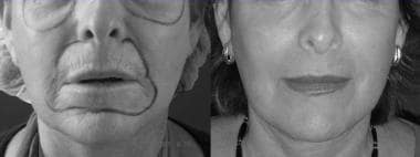
Before and after images are shown below.
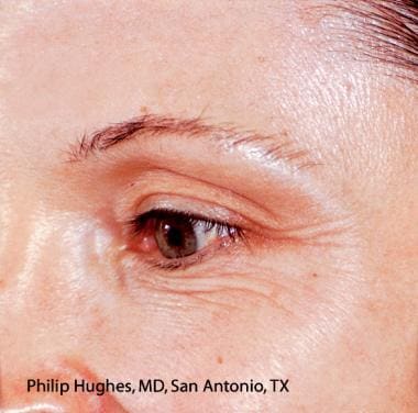 Before resurfacing with CB Erbium 2.94 laser. Phillip Hughes, MD, courtesy of HOYA ConBio.
 Same patient as in image above. After resurfacing with CB Erbium 2.94 laser. Phillip Hughes, MD, courtesy of HOYA ConBio.
 Before resurfacing with CB Erbium 2.94 laser. David Vasily, MD, courtesy of HOYA ConBio.
 Same patient as in image above. After resurfacing with CB Erbium 2.94 laser. David Vasily, MD, courtesy of HOYA ConBio.
 Before (Left) and after (right) upper and lower lip resurfacing with Erbium 2.94 laser. Courtesy of Meir Cohen, MD.
A study by Sayan et al of 547 patients who underwent field- or lesion-directed Er:YAG laser resurfacing for seborrheic or actinic keratosis found that over a 12-month review period, keratosis did not recur in any of the individuals treated with field-directed therapy, compared with six patients managed with lesion-directed resurfacing. The investigators also found the incidence of actinic keratosis recurrence with malignant transformation to be about 2.5%. [55]
Future and Controversies
The versatility of the Er:YAG lasers suggests several potential modifications of the original operative technique. [56] The laser can be used to ablate the epidermis, and then it can be reset to coagulate within the dermis at various levels to stimulate more collagen formation. (A study by El-Domyati et al found that fractional Er:YAG laser therapy for upper facial rejuvenation stimulated new collagen formation [types I, III, and VII] up to 6 months posttreatment. [57] ) The optimal depth within the dermis remains to be determined.
Another use under investigation is to ablate residual thermal damage after carbon dioxide laser resurfacing or even after deep Er:YAG lasing; removal of the proinflammatory coagulated tissue could result in less erythema and faster healing, but the tissue removed could be needed to stimulate new collagen formation. Preliminary work suggests that no significant difference is noted in the amount of collagen injury, inflammation, and new collagen formation between various combinations of the Er:YAG and carbon dioxide lasers soon after treatment. [1, 6, 58]
New Er:YAG lasers are being tested that produce high fluence values and smaller pulse durations, which, when used with a cryogen to cool the epidermis, allow the laser energy to spare the epidermis and only ablate and coagulate the dermis. Subepidermal laser resurfacing is attractive to patients who do not have a significant amount of photodamage because it reduces the erythema and edema associated with laser resurfacing. These nonablative techniques were introduced with Nd:YAG lasers and intense pulsed light systems, which are currently challenging the market share of the Er:YAG lasers. [59]
The latest innovation is the aforementioned fractional photothermolysis with Er:YAG laser (eg, PROfractional [Sciton Inc., Palo Alto, Calif], Pixel [Alma Lasers Ltd, Caesarea, Israel], Fraxel [Reliant Technologies, Mountain View, Calif]). This technique is based on creating spatially precise microscopic thermal wounds. The wounds created by the laser are narrow, sharply defined columns of skin known as microscopic thermal zones. According to preliminary reports, this resurfacing technique has been shown to be both safe and effective for improving facial and nonfacial photodamage, atrophic acne scars, hypopigmented scars, and dyspigmentation. [60, 61] Healing time is 3-6 days. Because only a fraction of the skin is treated during a single session, a series (typically, 4 treatments) of fractional resurfacing at 4-wk intervals is required for the best clinical improvement. [60, 61, 62]
A study by Cohen and Ross indicated that in patients undergoing facial rejuvenation, treatment combining fractional ablative 2940-nm and nonablative 1440-nm lasers has similar effectiveness as, but fewer immediate side effects than, treatment with only ablative lasers. Combined treatments in the study used a fractional nonablative 1440-nm neodynium YAG laser and a fractional ablative 2940-nm Er:YAG laser, while purely ablative treatments employed a combined confluent/fractional ablative Er:YAG laser or a fractional ablative CO2 laser. [63]
The new laser resurfacing techniques, while becoming more popular because of easier recovery, may fall into disfavor if the results are not long-lasting and if the complications of scarring increase. The results of ongoing and future clinical trials will establish the relative effectiveness and indications of the Er:YAG and other types of lasers.
Outward beauty is not enough, and the woman who would appear fair must not be content with any common manner. Words, wit, play, sweet talk and laughter, surpass the work of too simple nature. —Pliny the Elder
-
Before resurfacing with CB Erbium 2.94 laser. Phillip Hughes, MD, courtesy of HOYA ConBio.
-
Same patient as in image above. After resurfacing with CB Erbium 2.94 laser. Phillip Hughes, MD, courtesy of HOYA ConBio.
-
Before resurfacing with CB Erbium 2.94 laser. David Vasily, MD, courtesy of HOYA ConBio.
-
Same patient as in image above. After resurfacing with CB Erbium 2.94 laser. David Vasily, MD, courtesy of HOYA ConBio.
-
Before (Left) and after (right) upper and lower lip resurfacing with Erbium 2.94 laser. Courtesy of Meir Cohen, MD.