Article

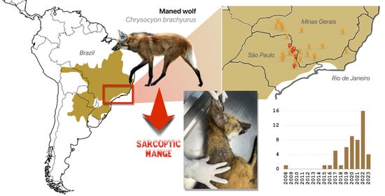
The Sarcoptic Mange in Maned Wolf (Chrysocyon brachyurus): Mapping an Emerging Disease in the Largest South American Canid

by
Flávia Fiori
1,2,
Rogério Cunha de Paula
1,3,
Pedro Enrique Navas-Suárez
4,
Ricardo Luiz Pires Boulhosa
1 and
Ricardo Augusto Dias
2,*
1
Instituto Pró-Carnívoros, Atibaia 12945-010, Brazil
2
Department of Preventive Veterinary Medicine and Animal Health, School of Veterinary Medicine, University of Sao Paulo, São Paulo 05508-270, Brazil
3
Centro Nacional de Pesquisa e Conservação de Mamíferos Carnívoros-CENAP, Instituto Chico Mendes para a Conservação da Biodiversidade—ICMBio, Ministry of Environment and Climate Change, Atibaia 12952-011, Brazil
4
Department of Pathology, School of Veterinary Medicine, University of Sao Paulo, São Paulo 05508-270, Brazil
*
Author to whom correspondence should be addressed.
Pathogens 2023, 12(6), 830; https://doi.org/10.3390/pathogens12060830
Submission received: 16 May 2023 / Revised: 13 June 2023 / Accepted: 14 June 2023 / Published: 15 June 2023
(This article belongs to the Special Issue Emerging Infections in Domestic Animals)

Abstract

:
The maned wolf (Chrysocyon brachyurus) is the largest South American canid. In Brazil, as in other countries, it is considered an endangered species. Habitat loss, landscape changes, hunting, and roadkill are the main threats to this species. In addition, invasive diseases of domestic animals are considered to be an emerging threat to the maned wolf, where parasitic diseases are relevant. Sarcoptic mange is a skin disease caused by the mite Sarcoptes scabiei. This disease is currently almost globally distributed, with a remarkable host diversity. In Brazil, reports of sarcoptic mange in wildlife include several species, both wild and captive. However, the impact of this disease on wildlife is unknown. At the time of writing, there is only one published report of sarcoptic mange in maned wolves. This study sheds light on the occurrence of sarcoptic mange in free-ranging maned wolves in their natural range. A total of 52 cases (suspected and confirmed) of sarcoptic mange were identified through social media review, camera trapping, chemical immobilization and sample collection. These cases were distributed in southeastern Brazil, in the states of São Paulo (n = 34), Minas Gerais (n = 17), and Rio de Janeiro (n = 1), demonstrating a rapid and widespread spread of this disease, although it still only occurs in part of the species’ range. We expect that these results will help to subsidize future actions relevant to the control of this emerging disease.

Graphical Abstract

1. Introduction

The maned wolf (Chrysocyon brachyurus) is the largest native South American canid, weighing up to 30 kg and inhabiting the central part of South America, especially the Cerrado, Chaco and Pampa biomes. Its diet consists primarily of the fruits of a typical savanna shrub, the wolf apple (Solanum lycocarpum), and secondarily, of rodents, birds [1], and invertebrates [2]. Despite this, individuals exhibit a dispersed social system, i.e., are essentially solitary, which is maintained by avoidance (barking), visual threat displays or fecal pheromones [2]. Adult home ranges vary across the distribution area but in natural habitats can be as large as around 80 km2, with little overlap with the home ranges of not paired individuals [3,4]. Mating pairs eventually locate close to each other simultaneously in the fall, but maintain a distance of >0.5 km from each other, regardless of the time of the day [3]. Publications on interspecific interactions with vertebrates other than their prey are scarce.
In Brazil, this species is classified as vulnerable (VU) [4]. Globally, however, it is classified as near threatened (NT) [5]. Habitat loss and continuous landscape changes due to agriculture and ranching, mining and other human activities, road mortality, retaliatory hunting (as a result of predation of domestic birds), and transmissible diseases are the main threats to the species [4]. Currently, transmissible diseases play an important role in the decline of carnivore populations, and are considered a major threat [6,7].
Direct contact between pets and wildlife is known to promote the bi-directional flow of pathogens [8]. The impact of domestic animal pathogens on wildlife is still unclear. However, some examples demonstrate their detrimental effects (morbidity and even mortality), such as canine distemper (CDV) outbreaks in African wild dogs in the Serengeti [9], and in the Mexican wolves in the USA [10]. There are also increasing reports of endoparasites such as Dirofilaria imitis [11], Toxocara canis [12], and Tricuris vulpis [13] in wild canids. Several pathogens have been reported in the maned wolf, some of which are of domestic origin [14,15,16,17,18,19,20,21,22,23,24].
Sarcoptic mange is a parasitic disease caused by the mite Sarcoptes scabiei. It is almost globally distributed, and has been described in humans and animals (domestic and wild) [25,26]. In Brazil, sarcoptic mange has been reported in wild [27,28,29,30] and captive animals [31]. However, despite its wide (geographic and species) distribution, reports of sarcoptic mange in Brazilian wild carnivores are rare. Sarcoptic mange has been described in humans since the mid-13th century and only later, in the 19th century, in livestock [32]. The entire life cycle of the ectoparasite occurs within the host: the females form burrows in the stratum corneum of the skin, then emerge to mate on the surface and lay eggs, which take 3 to 4 days to hatch [33]. The presence of females in the skin’s stratum corneum and their secretions cause a hypersensitivity reaction in the host, resulting in severe pruritus [34]. Depending on the development of clinical signs, the host may die from secondary infections and/or cachexia [35].
Sarcoptic mange has been described in crab-eating fox (Cerdocyon thous) [29,30,36], chilla fox (Lycalopex griseus) [37], sechuran fox (L. sechurae) [38], bush dog (Speothos venaticus) [39,40], and maned wolf (C. brachyurus) in Bolivia [41] and Brazil [39]. However, all reports are case descriptions. Prior to the present work, there is only one published report of sarcoptic mange in the maned wolf [41]. Nevertheless, the aim of this study was to collect information on the occurrence of sarcoptic mange in free-ranging maned wolves in their natural range in Brazil.

2. Methodology

2.1. Study Design, Study Area and Study Period

This study included confirmed cases (group G1, hereafter G1) and suspected cases (group G2, hereafter G2). G1 individuals were captured with box traps or rescued from September 2017 to February 2023. In addition, we included in this group a record traced back to January 2008 (the first record of sarcoptic mange in maned wolves, to our knowledge [41]). For the G2, camera trap images were obtained (G2A) and an active social media search was conducted (G2B). The increasing number of confirmed cases (G1) motivated the expansion of the search for new potential cases (G2). G2A individuals were enrolled from December 2016 to February 2023, and G2B individuals were enrolled from October 2017 to March 2023. G1 and G2A animals were monitored by the Pró-Carnívoros Institute and the National Research Center for Carnivore Conservation (CENAP) of the Chico Mendes Institute for the Biodiversity Conservation (Brazilian Ministry of Environment and Climate Change). All the data collected were in accordance with the actions of the National Action Plan for Wild Canids (https://www.gov.br/icmbio/pt-br/assuntos/biodiversidade/pan/pan-canideos (accessed on 16 of January 2023)). The study area for G1 and G2A included the state of São Paulo, along the border with the state of Minas Gerais, in southeastern Brazil (Figure 1). For G1, 14 sampling points were monitored and for G2A, 49, according to the activities of the “Lobos do Pardo” project and indications of suitable habitats for the presence of maned wolves. Records for G2B were obtained elsewhere in the maned wolf range, in Brazil. The maned wolf’s range was obtained in the IUCN Spatial Data Download (https://www.iucnredlist.org/resources/spatial-data-download (accessed on 17 of January 2023)) and all cases were plotted on a thematic map prepared in the QGIS version 3.20 (Odense) software.

2.2. Confirmed Cases (Group G1)

Since there are no pathognomonic lesions of sarcoptic mange, a case was considered confirmed if mites were identified in skin scrapings or biopsies. One case included in this study was previously described by Jorge and Jorge [39].
The number of box traps used simultaneously in each sampling site varied from 2 to 6 (mean = 4). The box traps were used for physical containment and were distributed over the monitored sampling site at pre-determined locations. A dose of 4 mg/kg of Zoletil 50® (tiletamine hydrochloride 125.0 mg and zolazepam hydrochloride 125.0 mg) was used for chemical containment. The anesthetic was administered intramuscularly, using a hypodermic syringe. Clinical parameters (heart rate, respiratory rate, rectal temperature, saturation, and eyelid, anal, and digital reflexes) were monitored throughout the procedure. A general condition assessment was performed after chemical containment, and age was estimated based on the dental arches [42,43,44]. Weight was assessed and compared to a scale proposed by Emmons [45] and ICMBio [46]. Animals were fully examined for skin lesions and deep skin scrapings, and punch biopsies were collected under chemical restraint. The scrapings were placed on slides and later examined under a light microscope to look for mites. For histopathologic examination, the skin fragments were fixed in 10% neutral buffered formalin, embedded in paraffin wax (FFPE), sectioned at 5 μm, and stained with hematoxylin and eosin for light microscopy. Morphological identification of Acari in tissue sections was based on published guidelines [47]. Captured wolves were monitored with radio collars throughout the study period. After anesthetic recovery, the animals were released at the same capture site.

2.3. Suspected Cases (Group G2)

For this assessment, we use camera trap surveys (G2A) and social media posts (G2B) containing descriptions or photographs of maned wolves with suggestive mange skin lesions. For G2A, a total of 71 different sites were used for camera trapping during the study, with the number of simultaneous sites varying depending on the season and the duration of each field campaign. A number of 30–35 cameras were left in fixed positions throughout the entire sampling period and 15–20 cameras were repositioned after one year. In the daytime images captured by camera traps, alopecia could be identified from the frames of the recorded videos. For the night images, alopecia was observed as peculiar dark patches on the exposed skin in contrast to the animal’s fur.
For G2B, we considered records whose published images contained one or more clinical signs of the disease, such as alopecia, scabbing, desquamation, and skin lesions (due to pruritus) at the elbows, hocks, and/or margin of the pinna. The authors of the social media posts (from governmental institutions, NGOs, veterinarians or biologists) were contacted and asked if, in addition to the suggestive clinical signs, pruritus was observed, as well as the exact location of the recording.
In addition, if identification of the mite in skin scrapings or biopsies from the captured wolves (G1) was not possible, and only the presence of the suggestive lesions was observed, these animals were included in G2A.

3. Results

A total of 52 cases of sarcoptic mange were identified, of which 10 (19.2%) were confirmed (G1) and 42 (80.8%) were suspected (Table 1, Figure 1), including 12 (28.6%) from G2A and 30 (71.4%) from G2B. According to the origin, 34 (65.4%) cases were from the state of São Paulo (including all the animals from G1, in addition to 11 of animals from G2A and 13 from G2B), 17 cases from the state of Minas Gerais (one from G2A and 16 from G2B) and one case from the state of Rio de Janeiro (G2B). Figure 2 shows examples of day and night images with the characteristic signs of sarcoptic mange.
Of the confirmed animals (G1), eight (53%) were males and seven (47%) were females. The body condition of 13 (86.7%) animals was within the normal body weight range for the species, including 3 juveniles (Table S1—Supplementary Material). In six cases, mites were seen in the biopsies, and in four animals, the deep skin scraping was positive. Of the 42 animals in G2, 7 (16.7%) were captured with camera traps in the study area, and in 5, no mites were observed in skin scrapings and biopsies (11.9%) (G2A), and 30 (71.4%) were collected indirect data collection (G2B).
According to the period in which records were noticed, considering G1 and G2: in 2008, one case; from 2009 to 2014, no record was made; in 2015, one case; in 2016, one case; in 2017, five cases; and in 2018, one case. The records became abundant from 2019 on, with six cases; in 2020, nine cases; in 2021, eight cases; in 2022, sixteen cases; and in 2023, four cases were recorded by March.
The trapping effort (G1) was 18,312 traps × hours. A total of 67 captures of 20 individual maned wolves occurred at all 14 monitoring sites. The sampling effort for the camera trap survey (G2A) was 870,168 traps × hours, resulting in 731 images of 43 individual maned wolves (adult and juveniles), at 27 sampling sites (55% of the monitored sampling sites).
Microscopically, in three cases, the epidermis showed marked parakeratotic hyperkeratosis, degenerate and non-degenerate neutrophils, and multifocal areas of ulceration associated with numerous intralesional adult mites. Within the dermis, a mild mixed inflammatory infiltrate (eosinophils, neutrophils, plasma cells with fewer macrophages and lymphocytes), congestion of deep dermal vessels, and lymphangiectasia were observed. Morphologically, the mites measure between 200 × 400 μm, have articulated appendages, a chitinous exoskeleton, dorsal spines, striated muscle, intestinal and reproductive structures, and a body cavity (Figure 2).

4. Discussion

The present study described the occurrence of sarcoptic mange in maned wolves in southeastern Brazil. The methodology was limited by the small number of skin scrapings and biopsies obtained from affected animals, which prevented a comprehensive understanding of the dermatological manifestations and maintenance of the disease in this species. Despite these limitations, it is important to emphasize the commendable sampling effort undertaken in this study, which allowed the collection of valuable data on sarcoptic mange in maned wolves.
The increasing number of individuals affected by sarcoptic mange reported in a small portion of the maned wolf’s range in Brazil (approximately 6% of the total species’ total range in the country), suggests that this disease is a new emerging threat to the species, especially in anthropized areas. The first report of sarcoptic mange in a maned wolf in Brazil was recorded in 2008 [39]. In this study, all possible sources of information were searched for suspected or confirmed cases of sarcoptic mange in maned wolves throughout the species range. From 2008 to 2019, only a few records of animals with clinical signs of sarcoptic mange were registered. The vast majority of cases were recorded in the study area after 2019. With the exception of the records from the states of Minas Gerais (Cbr 33, 38) and Rio de Janeiro (Cbr 42), the recorded cases from the state of São Paulo represent the core zone of the disease circulation. In contrast to maned wolves, records of sarcoptic mange in other wild canids, such as crab-eating foxes and bush dogs, are distributed throughout their range [29,30,36,39,40].
Observations in G1 animals revealed areas of alopecia and crusts on the skin as clinical signs. However, skin samples from five animals with clinical signs were negative in the parasitological and histopathological diagnoses, and were included in the G2A group. Although a proper protocol for mite isolation has not been established, it has been suggested that the collection of mites depends on the stage of infestation, as they may be difficult to collect at certain stages, leading to false negative results in the laboratory techniques used [35,48]. Thus, our results suggest that animals in the early and late stages of infestation are diagnosed by skin scraping. On the other hand, animals in the intermediate stage of the infestation are diagnosed by skin biopsy for histopathologic analysis.
Individuals Cbr 5, Cbr 8 and Cbr 9 (which were negative in skin scraping) all had a history of direct (pairs during breeding season or parents with youngsters) and/or indirect contact (high home range overlap) with other wolves positive for sarcoptic mange (Cbr 6, 7, 14 and Cbr 10, 11, 12, respectively). This contact was recorded using GPS collar monitoring installed on the animals. Once it was known that the mange transmission occurs either directly, through physical contact, or indirectly, through fomites [49,50], clinical diagnosis was applied to these animals. Both wolves (Cbr 5 and Cbr 9) showed characteristic clinical signs of sarcoptic mange such as alopecia, scaling, and crusting of the skin, in addition to clinical cure after in loco treatment with Bravecto® (25–65 mg of fluralaner/kg of body weight). Although these animals showed improvement in the clinical signs, the diagnosis of sarcoptic mange could not be confirmed because Bravecto® (Merck & Co., Inc., Rahway, NJ, USA) does not specifically target Sarcoptes scabiei.
Animals included in the G2 group were classified as suspect because the images showed clinical signs suggestive of sarcoptic mange [26,35]. Signs such as alopecia, crusts, and scaling (the latter two clinical signs were only visible in the daytime images) were observed in camera trap images. As the camera traps were used for a long-term survey in the study sites, the individuals included in the study could be registered over several months, allowing the analysis of the evolution of the infestation process. Thus, the observations from different stages of infestation of the animals in groups G1 and G2A served as a basis for comparing the clinical status of the wolves included in G2B. G2B animals were separated spatially and temporally, reducing the likelihood of duplicate cases. In addition, the maned wolf is not a gregarious species and therefore does not form permanent groups. For this reason, relationships between individuals living in the same groups or not and passing the mite among themselves are unlikely.
All cases of sarcoptic mange (G1 or G2) were recorded in areas where the landscape is changing due various human activities, especially agriculture and cattle ranching [51]. In addition, camera trap data show an overlap in the land use between maned wolves (groups G1 and G2A) and domestic animals such as dogs and cattle, two species commonly affected by sarcoptic mange [52]. Indirect contact between wild and domestic canids in all study areas may suggest the potential transmission of the mites through the modified areas. However, at the other study sites in other parts of Brazil, camera trapping also revealed that maned wolves and domestic animals shared anthropized areas, and yet there are no records of infested wild canids, except for crab eating foxes and sometimes bush dogs. In addition, according to Sinclair et al. [53], environment change due to climate change, the movement and migration of hosts, and the presence of new pathogens may bring both domestic and wild species into closer contact in areas where this would not have been possible. This process favors the emergence of infectious diseases [53].
The possibility of anthropogenic changes influencing the epidemiology of sarcoptic mange is suggested. As with domestic dogs and cattle, there are other species that could be part of the epidemiological cycle of mange. Exotic species may be affected and transmit the parasites. Examples include the wild boar (Sus scrofa), which has been reported to have mange in its natural range [54,55], and lagomorphs such as the European hare (Lepus europaeus), which, although not reported to have mange, has mange in other members of the genus [56]. These exotic species are found in different regions of Brazil, in sympatry with the maned wolf [57,58]. Therefore, epidemiologic surveillance is proposed for these four domestic and exotic species, considering their contribution to the epidemiology of sarcoptic mange.

5. Concluding Remarks

The records compiled in this study and the mapping of maned wolf sarcoptic mange are a first step in further evaluating how an emerging disease may expand its range in relation to variables related to human’s presence. The information generated leads to a negative alert regarding a high impact on the health of an endangered species. This emerging disease must be a high priority level for maned wolf conservation strategies. Once it has been established that the epidemiology of sarcoptic mange can be linked to landscape and bioclimatic variables, as well as to close contact with invasive and domestic species, preventive strategies must be put into practice; nevertheless, genetic studies of mite populations must be carried out to elucidate the transmission chain of sarcoptic mange involving maned wolves and other hosts, whether domestic (e.g., dogs) or wild, whether invasive or native. In particular, educational efforts focused on proper land use and responsible care of the domestic animals are important issues to be developed for the conservation of the maned wolf. Disease surveillance of the Brazilian fauna is gradually being standardized and professionalized. In the case of the native wild canids, these actions are being discussed in the National Action Plan for Wild Canids of the Instituto Chico Mendes de Conservação da Biodiversidade (ICMBio) of the Brazilian Ministry of Environment and Climate Change, and the results will be useful in prioritizing public policies to help reduce the spread of sarcoptic mange in maned wolves.

Supplementary Materials

The following supporting information can be downloaded at: https://www.mdpi.com/article/10.3390/pathogens12060830/s1, Table S1: General data of captured wolves and sarcoptic mange diagnosis.

Author Contributions

Study design: F.F., R.C.d.P. and R.A.D.; Data collection: F.F., R.C.d.P. and R.L.P.B.; Data Analysis: F.F., R.C.d.P., P.E.N.-S. and R.A.D.; Writing: F.F., R.C.d.P., P.E.N.-S. and R.A.D.; Revision: F.F., R.C.d.P., P.E.N.-S., R.L.P.B. and R.A.D. All authors have read and agreed to the published version of the manuscript.

Funding

This research was funded by ICMBio (2018-2023), AES Tietê (Lobos do Pardo Project 2018-2020), Ideal Wild, CAPES (Finance Code 001).

Institutional Review Board Statement

All research protocols were approved by SISBIO (Sistema de Autorização e Informação em Biodiversidade)/ICMBio (licenses 61097, 830092), and the Animal Ethical Committee (CEUA—USP) under license registration 8983240322.

Informed Consent Statement

Not applicable.

Data Availability Statement

Data available on request due to restrictions.

Acknowledgments

The authors acknowledge the farms that allow the animal captures and data collection. Specifically, Fazenda Paineiras, Fazenda Belo Monte, Fazenda Jequitibá, Fazenda da Serra, Fazenda da Mata and Fazenda Ambiental Fortaleza. The companies and institutions AES Brasil, Parc Animalier de La Barben, Zoo la Bourbansais, Idea Wild, Log Nature, Fundação Florestal, Environmental Police of Itatiba, Tambaú and Andradas, and LAPCOM-VPT-FMVZ-USP. Yet, Jean Pierre Santos, Joares May Jr., Christiane Rieken, Maria Fernanda Gondim, Helia Piedade, Márcio Siconelli, Jeferson Pires, Bruno Troiano and Project Tatu Canastra for sample collection and/or sharing information. To Coordination of Superior Level Staff Improvement—CAPES (Finance Code 001).

Conflicts of Interest

The authors declare no conflict of interest.

References

  1. Motta-Junior, J.C.; Talamoni, S.A.; Lombardi, J.A.; Simokomaki, K. Diet of the maned wolf, Chrysocyon brachyurus, in central Brazil. J. Zool. 1996, 240, 277–284. [Google Scholar] [CrossRef]
  2. Kleiman, D.G. Social Behavior of the Maned Wolf (Chrysocyon brachyurus) and Bush Dog (Speothos venaticus): A Study in Contrast. J. Mammal. 1972, 53, 791–806. [Google Scholar] [CrossRef]
  3. Jácomo, A.T.A.; Kashivakura, C.K.; Ferro, C.; Furtado, M.M.; Astete, S.P.; Tôrres, N.M.; Sollmann, R.; Silveira, L. Home Range and Spatial Organization of Maned Wolves in the Brazilian Grasslands. J. Mammal. 2009, 90, 150–157. [Google Scholar] [CrossRef]
  4. Paula, R.C.; Rodrigues, F.H.G.; Queirolo, D.; Jorge, R.P.S.; Lemos, F.G.; Rodrigues, L.A. Avaliação do estado de conservação do lobo-guará Chrysocyon brachyurus (Illiger, 1815) no Brasil. Biodivers. Bras. 2013, 3, 146–159. [Google Scholar]
  5. Paula, R.C.; Dematteo, K. Chrysocyon brachyurus. In The IUCN Red List of Threatened Species; 2015; Available online: https://www.iucnredlist.org/species/4819/88135664 (accessed on 1 December 2021). [CrossRef]
  6. Gittleman, J.L.; Funk, S.M.; MacDonald, D.W.; Wayne, R.K. (Eds.) Carnivore Conservation; Cambridge University Press: Cambridge, UK, 2001; Volume 5. [Google Scholar]
  7. May, J.A., Jr.; Felippe, P.A.N. Conservation Medicine. In Ecology and Conservation of the Maned Wolf—Multidisciplinary Perspectives; Consorte-McCrea, A.G., Santos, E.F., Eds.; CRC Press: Boca Raton, FL, USA, 2014. [Google Scholar]
  8. Miller, R.S.; Farnsworth, M.L.; Malmberg, J.L. Diseases at the livestock–wildlife interface: Status, challenges, and opportunities in the United States. Prev. Vet. Med. 2013, 110, 119–132. [Google Scholar] [CrossRef] [PubMed] [Green Version]
  9. Goller, K.V.; Fyumagwa, R.D.; Nikolin, V.; East, M.L.; Kilewo, M.; Speck, S.; Müller, T.; Matzke, M.; Wibbelt, B. Fatal canine distemper infection in a pack of African wild dogs in the Serengeti ecosystem, Tanzania. Vet. Microbiol. 2010, 146, 245–252. [Google Scholar] [CrossRef] [PubMed] [Green Version]
  10. Justice-Allen, A.; Clement, M.J. Effect of Canine Parvovirus and Canine Distemper Virus on the Mexican Wolf (Canis lupus baileyi) Population in the USA. J. Wildl. Dis. 2019, 55, 682–688. [Google Scholar] [CrossRef]
  11. Penezić, A.; Selakovic, S.; Pavlović, I.; Ćirović, D. First findings and prevalence of adult heartworms (Dirofilaria immitis) in wild carnivores from Serbia. Parasitol. Res. 2014, 113, 3281–3285. [Google Scholar] [CrossRef]
  12. Okulewicz, A.; Perec-Matysiak, A.; Buńkowska, K.; Hildebrand, J. Toxocara canis, Toxocara cati and Toxascaris leonina in wild and domestic carnivores. Helminthologia 2012, 49, 3–10. [Google Scholar] [CrossRef] [Green Version]
  13. Criado-Fornelio, A.; Gutierrez-Garcia, L.; Rodriguez-Caabeiro, F.; Reus-Garcia, E.; Roldan-Soriano, M.; Diaz-Sanchez, M. A parasitological survey of wild red foxes (Vulpes vulpes) from the province of Guadalajara, Spain. Vet. Parasitol. 2000, 92, 245–251. [Google Scholar] [CrossRef]
  14. Dietz, J.M. Ecology and social organization of the maned wolf (Chrysocyon brachyurus). In Smithsonian Contributions to Zoology, n. 392; Smithsonian Institution Press: Washington, DC, USA, 1984; pp. 1–51. [Google Scholar] [CrossRef]
  15. Deem, S.L.; Emmons, L.H. Exposure of free-ranging maned wolves (Chrysocyon brachyurus) to infectious and parasitic disease agents in the Noël Kempff Mercado National Park, Bolivia. J. Zoo Wildl. Med. 2005, 36, 192–197. [Google Scholar] [CrossRef] [Green Version]
  16. Provença, L.M. Soroprevalência de Doenças Infecciosas Caninas em Populações de Lobo-Guará (Chrysocyon brachyurus) e Cachorro-do-Mato (Cerdocyon thous) na Estação Ecológica de Águas Emendadas, DF. Master’s Thesis, Universidade de Brasilia, Brasília, Brazil, 2007. [Google Scholar]
  17. Vieira, F.M.; Luque, J.; Muniz-Pereira, L.C. Checklist of helminth parasites in wild carnivore mammals from Brazil. Zootaxa 2008, 1721, 1–23. [Google Scholar] [CrossRef]
  18. Braga, R.T.; Vynne, C.; Loyola, R.D. Fauna parasitária intestinal de Chrysocyon brachyurus (lobo-guará) no Parque Nacional das Emas. Bioikos 2010, 24, 49–55. [Google Scholar]
  19. Mattos, P.S.R. Epidemiologia e Genética Populacional do Lobo-Guará, Chrysocyon brachyurus (Illiger, 1915) (Carnívora, Canidae) na Região Nordeste do Estado de São Paulo. Ph.D. Thesis, Universidade Federal de São Carlos, São Carlos, Brazil, 2003. [Google Scholar]
  20. Braga, R.T.; Vynne, C.; Corrêa, M.C.R.; Loyola, R.D. New record of Dyoctophyma renale in the maned wolf (Chrysocyon brachyrus) in the state of Goiás, Brazil. Bioikos 2010, 24, c43–c47. [Google Scholar]
  21. Vieira, F.M. Helmintos Parasitos de Mamíferos Carnívoros Silvestres no Município de Juiz de Fora, Zona da Mata do Estado de Minas Gerais, Brasil. Ph. D. Thesis, Universidade Federal Rural do Rio de Janeiro, Rio de Janeiro, Brazil, 2011. [Google Scholar]
  22. Arrais, R.C.; Paula, R.C.; Martins, T.F.; Nieri-Bastos, F.A.; Marcili, A.; Labruna, M.B. Survey of ticks and tick-borne agents in maned wolves (Chrysocyon brachyurus) from a natural landscape in Brazil. Ticks Tick-borne Dis. 2021, 12, 101639. [Google Scholar] [CrossRef]
  23. Rodrigues, T.O.; Canelo, E.A.; Sommerfeld, S.; Moraes, F.P.; Nascimento, F.G.O.; Silva, D.M.; Lima, A.M.C.; Santos, A.L.Q. Cinomose em lobo-guará Chrysocyon brachyurus (Illiger, 1811): Relato de caso. Veterinária Notícias 2014, 20, 24. [Google Scholar]
  24. Vieira, E.G.; Araújo, G.V.B.; Tolomeu, A.L.G.; Cardoso, V.A.F.X.; Mendes, A.L.P.; Machado, J.P. Infecção por Dioctophyma renale com localização livre em cavidade abdominal de lobo-guará (Chrysocyon brachyurus): Relato de caso. In Proceedings of the VI Simpósio de Produção Acadêmica (SIMPAC 2014), Viçosa, Brazil, 22–28 October 2014; pp. 363–368. [Google Scholar]
  25. Escobar, L.E.; Carver, S.; Cross, P.C.; Rossi, L.; Almberg, E.S.; Yabsley, M.J.; Niedringhaus, K.D.; Van Wick, P.; Dominguez-Villegas, E.; Gakuya, F.; et al. Sarcoptic mange: An emerging panzootic in wildlife. Transbound. Emerg. Dis. 2022, 69, 927–942. [Google Scholar] [CrossRef]
  26. Astorga, F.; Carver, S.; Almberg, E.S.; Sousa, G.R.; Wingfield, K.; Niedringhaus, K.D.; Van Wick, P.; Rossi, L.; Cross, P.; Angelone, S.; et al. International meeting on sarcoptic mange in wildlife, June 2018, Blacksburg, Virginia, USA. Parasites Vectors 2018, 11, 449. [Google Scholar] [CrossRef] [PubMed] [Green Version]
  27. Miranda, F. Cingulata (Tatus) e Pilosa (Preguiças e Tamanduás). In Tratado de Animais Selvagens Medicina Veterinária, 2nd ed.; Cubas, Z.S., Silva, J.C.R., Catão-Dias, J.L., Eds.; Roca: São Paulo, Brazil, 2014; Volume 1, pp. 707–722. [Google Scholar]
  28. Nogueira, M.F.; Cruz, T.F. Doenças da Capivara; Embrapa Pantanal: Corumbá, Brazil, 2007. [Google Scholar]
  29. Almeida, J.C.; Leal, C.A.S.; Melo, R.P.B.; Albuquerque, P.P.F.; Pedrosa, C.M.; Zermiani, F.C.; Farias, R.C.; Mota, R.A. Co-Infection by Sarcoptes scabiei and Microsporum gypseum in Free-Ranging Crab-Eating Fox, Cerdocyon thous (Linnaeus, 1766). Braz. Arch. Biol. Technol. 2018, 61, e18160508. [Google Scholar] [CrossRef]
  30. Teodoro, T.G.; Lima, P.A.; Stehling, P.C.; Junior, I.M.O.; Varaschin, M.S.; Wouters, F.; Wouters, A.T.B. Sarcoptic mange (Sarcoptes scabiei) in wild canids (Cerdocyon thous). Pesq. Vet. Bras. 2018, 38, 1444–1448. [Google Scholar] [CrossRef] [Green Version]
  31. Diniz, L.S.; Costa, E.O.; Benites, N.R. Processos dermatológicos em animais silvestres: Avaliação clínica e laboratorial de dermatopatias em silvestres revela prevalência de causas parasitárias, bacterianas e fúngicas. Clin. Vet. 1997, 2, 16–19. [Google Scholar]
  32. Currier, R.W.; Walton, S.F.; Currie, B.J. Scabies in animals and humans: History, evolutionary perspectives, and modern clinical management. Ann. N. Y. Acad. Sci. 2012, 1230, E50–E60. [Google Scholar] [CrossRef] [PubMed]
  33. Taylor, M.A.; Coop, R.L.; Wall, R.L. Parasitologia Veterinária; Guanabara: Rio de Janeiro, Brazil, 2017. [Google Scholar]
  34. Zachary, J.F.; McGavin, M.D. Bases da Patologia em Veterinária, 5th ed.; Elsevier: Rio de Janeiro, Brazil, 2013. [Google Scholar]
  35. Pence, D.B.; Ueckermann, E. Sarcoptic mange in wildilfe. Rev. Sci. Tech. OIE 2002, 21, 385–398. [Google Scholar] [CrossRef]
  36. Uzai, G.J.S.; Monteiro, C.P.; Soares, R.; Silva, M.A.; Oliveira, A.R.; Santos, R.; Nunes, L. Morphological and molecular diagnosis of diseases of free-ranging crab-eating foxes (Cerdocyon thous). Arq. Bras. Med. Vet. Zoo 2021, 73, 583–588. [Google Scholar] [CrossRef]
  37. Verdugo, C.; Espinoza, A.; Moroni, M.; Valderrama, R.; Hernandez, C. Sarcoptic Mange in a South American Gray Fox (Chilla Fox; Lycalopex griseus), Chile. J. Wildl. Dis. 2016, 52, 738–741. [Google Scholar] [CrossRef]
  38. Villalba-Briones, R.; Molineros, E.B.; Monrós, J.S. First report of Sarcoptes scabiei parasitism (Sarcoptiformes: Sarcoptidae) in Lycalopes sechurae (Mammalia: Carnivora). Rev. Bras. Parasitol. Vet. 2022, 31, e005022. [Google Scholar] [CrossRef] [PubMed]
  39. Jorge, R.S.P.; Jorge, M.L.S.P. Carnívora—Canidae (Cachorro-do-mato, Cachorro-vinagre, Lobo-guará e Raposa-do-campo). In Tratado de Animais Selvagens Medicina Veterinária, 2nd ed.; Cubas, Z.S., Silva, J.C.R., Catão-Dias, J.L., Eds.; Roca: São Paulo, Brazil, 2014; Volume 1, pp. 764–778. [Google Scholar]
  40. Jorge, R.S.P.; Lima, E.S.; Lucarts, L.E.B. Sarna sarcóptica ameaçando cachorros-vinagres (Speothos venaticus) de vida livre em Nova Xavantina, MT. In Proceedings of the XXXIII Congresso Anual Sociedade de Zoológicos do Brasil, Sorocaba, Brazil, 21–25 March 2009. [Google Scholar]
  41. Luque, J.A.D.; Muller, H.; González, L.; Berkunsky, I. Clinical sings suggestive of mange infestation in a free-ranging maned wolf (Chrysocyon brachyurus) in the Moxos Savannahs of Bene, Bolivia. Mastozool Neotrop 2014, 21, 135–138. [Google Scholar]
  42. Olifiers, N.; Bianchi, R.d.C.; D’Andrea, P.S.; Mourão, G.; Gompper, M.E. Estimating age of carnivores from the Pantanal region of Brazil. Wildl. Biol. 2010, 16, 389–399. [Google Scholar] [CrossRef] [Green Version]
  43. Chevallier, C.; Gauthier, G.; Berteaux, D. Age Estimation of Live Arctic Foxes Vulpes lagopus Based on Teeth Condition. Wildl. Biol. 2017, 2017, 1–6. [Google Scholar] [CrossRef] [Green Version]
  44. Sharma, C.P.; Sharma, V.; Goyal, S.P.; Stevens, H.; Gupta, S.K. Age estimation of Tiger Panthera tigris (Linnaeus, 1758) and Lion Panthera leo (Linnaeus, 1758) (Mammalia: Carnivora: Felidae): Applicability of cementum annuli analysis method. J. Threat. Taxa 2022, 14, 21805–21810. [Google Scholar] [CrossRef]
  45. Emmons, L.H. Maned Wolves of Noel Kempff Mercado National Park; Smithsonian Contributions to Zoology, n. 639; Smithsonian Institution Press: Washington, DC, USA, 2012; pp. 1–35. [Google Scholar] [CrossRef] [Green Version]
  46. Instituto Chico Mendes de Conservação da Biodiversidade [ICMBio]. Sumário Executivo do Plano de Ação Nacional Para a Conservação do Lobo-Guará; Ministry of Environment and Climate Change: Brasília, Brazil, 2017. [Google Scholar]
  47. Gardiner, C.H.; Poynton, S.L. An Atlas of Metazoan Parasites in Animal Tissues; Armed Forces Institute of Pathology, American Registry of Pathology: Washington, DC, USA, 1999; ISBN 1881041492. [Google Scholar]
  48. Hnilica, K.A. Dermatologia de Pequenos Animais: Atlas Colorido e Guia Terapêutico, 3rd ed.; Elsevier: Rio de Janeiro, Brazil, 2012. [Google Scholar]
  49. Urquhart, G.M.; Armour, J.; Duncan, J.L.; Dunn, A.M.; Jennings, F.W. Parasitologia Veterinária, 2nd ed.; Guanabara Koogan: Rio de Janeiro, Brazil, 1998. [Google Scholar]
  50. Carneiro, V.O. Ocorrência da Sarna Sarcóptica em Cães Domiciliados no Bairro Vila Verde na Cidade de Tabatinga—AM; Graduation Final Work; Universidade do Estado do Amazonas: Tabatinga, Brazil, 2019. [Google Scholar]
  51. Souza, C.; Azevedo, T. MapBiomas General Handbook; MapBiomas: São Paulo, Brazil, 2017; pp. 1–23. [Google Scholar]
  52. Conceição, L.G.; Loures, F.H. Sistema tegumentar. In Patologia Veterinária, 2nd ed.; Santos, R.L., Alessi, A.C., Eds.; Roca: Rio de Janeiro, Brazil, 2016. [Google Scholar]
  53. Sinclair, A.R.E.; Fryxell, J.M.; Caughley, G. Wildlife Ecology Conservation and Management, 2nd ed.; Blackwell Publishing: Hoboken, NJ, USA, 2006. [Google Scholar]
  54. Haas, C.; Origgi, F.C.; Akdesir, E.; Linhares, M.B.; Giovannini, S.; Mavrot, F.; Casaubon, J.; Ryser-Degiorgis, M.-P. First detection of sarcoptic mange in free-ranging wild boar (Sus scrofa) in Switzerland. Schweiz. Arch. Tierheilkd. 2015, 157, 269–275. [Google Scholar] [CrossRef] [PubMed]
  55. Haas, C.; Origgi, F.C.; Rossi, S.; López-Olvera, J.R.; Rossi, L.; Castillo-Contreras, R.; Malmsten, A.; Dalin, A.-M.; Orusa, R.; Robetto, S.; et al. Serological survey in wild boar (Sus scrofa) in Switzerland and other European countries: Sarcoptes scabiei may be more widely distributed than previously thought. BMC Vet. Res. 2018, 14, 117. [Google Scholar] [CrossRef] [PubMed] [Green Version]
  56. Cardells, J.; Lizana, V.; Martí-Marco, A.; Lavín, S.; Velarde, R.; Rossi, L.; Moroni, B. First description of sarcoptic mange in an Iberian hare (Lepus granatensis). Curr. Res. Parasitol. Vector-Borne Dis. 2021, 1, 100021. [Google Scholar] [CrossRef] [PubMed]
  57. de Faria, G.M.M.; Rosa, C.A.; Corrêa, G.L.C.; Puertas, F.; Jiménez, K.M.O.; Perillo, L.N.; Hufnagel, L.; Leles, B.; de Paula, R.C.; Rodrigues, F.H.G.; et al. Geographic distribution of the European hare (Lepus europaeus) in Brazil and new records of occurrence for the Cerrado and Atlantic Forest biomes. Mammalia 2016, 80, 497–505. [Google Scholar] [CrossRef]
  58. Instituto Brasileiro do Meio Ambiente e dos Recursos Renováveis [IBAMA]. Relatório Sobre Áreas Prioritárias Para o Manejo de Javalis: Aspectos ambientais, Socioeconômicos e Sanitários; IBAMA: Brasília, Brasil, 2019. [Google Scholar]
Figure 1. Spatial distribution of confirmed and suspected cases of sarcoptic mange in maned wolves in Brazil.
Pathogens 12 00830 g001
Figure 2. (A). BTH 986. Skin. Numerous intralesional adult mites (asterisk). HE, 40× (B). BTH 986. Skin. Numerous intralesional adult mites. HE, 40× (B1) (Inset). Note a female specimen of Sarcoptes scabiei in the superficial epidermis. The mites have jointed appendages (black arrowhead), a chitinous exoskeleton (red arrow), ovary (red arrowhead), and striated muscle (black arrow). HE, 400×. (C). Nighttime image showing a maned wolf with dark spots in areas of the coat, indicating alopecia. (D). Daytime image showing a maned wolf with areas of alopecia, crusts and cracks in the skin.
Pathogens 12 00830 g002
Table 1. Sarcoptic mange cases in maned wolves, according to identification groups.
Identification GroupsIndividualsDate of RecordMunicipalityState
(mm/yyyy)
G1Cbr 105/2022MococaSão Paulo
Cbr 401/2008ItatibaSão Paulo
Cbr 607/2019MococaSão Paulo
Cbr 704/2021MococaSão Paulo
Cbr 1012/2017Mogi-GuaçuSão Paulo
Cbr 1112/2017Mogi-GuaçuSão Paulo
Cbr 1212/2017Mogi-GuaçuSão Paulo
Cbr 1304/2021São João da Boa VistaSão Paulo
Cbr 1409/2018São José do Rio PardoSão Paulo
Cbr 4802/2023CajuruSão Paulo
G2G2ACbr 206/2022MococaSão Paulo
Cbr 308/2022MococaSão Paulo
Cbr 503/2019MococaSão Paulo
Cbr 804/2021MococaSão Paulo
Cbr 909/2017Mogi-GuaçuSão Paulo
Cbr 1505/2022MococaSão Paulo
Cbr 1609/2021MococaSão Paulo
Cbr 1706/2020ItapiraSão Paulo
Cbr 1804/2020Mogi-GuaçuSão Paulo
Cbr 1911/2020Mogi-GuaçuSão Paulo
Cbr 2010/2019São José do Rio PardoSão Paulo
Cbr 2103/2020BotelhosMinas Gerais
G2BCbr 2210/2022ItirapinaSão Paulo
Cbr 2307/2020Santo Antônio do JardimSão Paulo
Cbr 2404/2022São Bento do SapucaíSão Paulo
Cbr 2505/2022São Bento do SapucaíSão Paulo
Cbr 2611/2021São Bento do SapucaíSão Paulo
Cbr 2712/2020São CarlosSão Paulo
Cbr 2803/2022São José do Rio PardoSão Paulo
Cbr 2903/2019Serra NegraSão Paulo
Cbr 3002/2021ArceburgoMinas Gerais
Cbr 3107/2022BotelhosMinas Gerais
Cbr 3204/2022Campos AltosMinas Gerais
Cbr 3303/2020Córrego NovoMinas Gerais
Cbr 3411/2022UberlândiaMinas Gerais
Cbr 3505/2016JardinésiaMinas Gerais
Cbr 3609/2020Pouso AltoMinas Gerais
Cbr 3703/2017Santana da VargemMinas Gerais
Cbr 3801/2021São João del ReiMinas Gerais
Cbr 3901/2019São Gonçalo do SapucaíMinas Gerais
Cbr 4008/2022Soledade de MinasMinas Gerais
Cbr 4107/2015 UberlândiaMinas Gerais
Cbr 4207/2022Santo Antônio de PáduaRio de Janeiro
Cbr 4307/2022UberabaMinas Gerais
Cbr 4407/2021UberabaMinas Gerais
Cbr 45 12/2022InconfidentesMinas Gerais
Cbr 4601/2023ItirapinaSão Paulo
Cbr 4710/2020Monte Alegre do SulSão Paulo
Cbr 4912/2022ItirapinaSão Paulo
Cbr 5001/2019PedregulhoSão Paulo
Cbr 5103/2023CampestreMinas Gerais
Cbr 5201/2023TambaúSão Paulo
Disclaimer/Publisher’s Note: The statements, opinions and data contained in all publications are solely those of the individual author(s) and contributor(s) and not of MDPI and/or the editor(s). MDPI and/or the editor(s) disclaim responsibility for any injury to people or property resulting from any ideas, methods, instructions or products referred to in the content.

